| Journal of Food Bioactives, ISSN 2637-8752 print, 2637-8779 online |
| Journal website www.isnff-jfb.com |
Original Research
Volume 35, September 2026, pages 46-56
Pyroglutamyl peptide–rich rice protein hydrolysate restores cecal N-acetyl amino acids in high-fat-diet–fed rats, outperforming short-chain fatty acids as early microbial markers
Satoshi Miyauchia, b, Shiro Kajiwarac, Tomoko Asaia, Yasutaka Shigemurab, Kenji Satoa, *
aDivision of Applied Biosciences, Graduate School of Agriculture, Kyoto University, Kitashirakawa Oiwake-cho, Sakyo-ku, Kyoto 606 8502, Japan
bDepartment of Food and Nutrition, Tokyo Kasei University, 1-18-1 Kaga, Itabashi-ku, Tokyo 173 8602, Japan
cBansyu chomiryo Co., Ltd., 948, Nozato, Himeji, Hyogo 670 0811, Japan
*Corresponding author: Kenji Sato, Division of Applied Biosciences, Graduate School of Agriculture, Kyoto University, Kitashirakawa Oiwake-cho, Sakyo-ku, Kyoto 606 8502, Japan. E-mail: kenjisato6213@gmail.com
DOI: 10.26599/JFB.2026.95035455
Received: June 12, 2026
Revised received & accepted: July 28, 2026
| Abstract | ▴Top |
Pyroglutamyl-leucine (pyroGlu-Leu) is a food-derived modified peptide known to modulate the gut microbiota. This study examined the effects of orally administered rice protein hydrolysate (RPH) containing pyroGlu-Leu in rats fed a 45% high-fat diet that induced mild dysbiosis without obesity or severe liver dysfunction. In addition to short-chain fatty acids (SCFAs), several N-acetyl amino acids (NAAAs) were detected in cecal contents. The high-fat diet significantly decreased both SCFAs and NAAAs, whereas administration of RPH or synthetic pyroGlu-Leu restored the levels of NAAAs despite not recovering those of SCFAs. RPH did not alter the overall gut microbial composition but modified specific bacterial taxa that correlated with NAAA levels. These results suggest that NAAAs are gut microbiota-derived metabolites that respond more sensitively to food-derived modulation of the gut microbiota than SCFAs, and may serve as early indicators of microbiota–host metabolic interactions.
Keywords: Peptide; Pyroglutamyl peptide; Microbiota; Short-chain fatty acid; N-Acetyl amino acid
| 1. Introduction | ▴Top |
Pyroglutamyl peptides, which are non-enzymatically generated from peptides containing N-terminal glutaminyl residue, are widely present in food protein hydrolysates and fermented foods (Ejima et al., 2018; Gazme et al., 2019; Kiyono et al., 2013; Nagao et al., 2024; Rahmadian et al., 2025; Sato et al., 1998, 2013; Shirako et al., 2020). Several pyroglutamyl di- and tri-peptides have been reported to resist in vitro digestion by endoproteinases and exopeptidases (Chen et al., 2019; Ejima et al., 2018; Miyauchi et al., 2022; Sato et al., 1998). In addition, levels of certain food-derived pyroglutamyl peptide increase in the bloodstream after oral administration of food protein hydrolysates and fermented foods, such as soybean paste (miso) and fish sauce (Ejima et al., 2018; Higaki-Sato et al., 2006; Miyauchi et al., 2022; Nagao et al., 2024; Rahmadian et al., 2025; Sato et al., 2013).
Several pyroglutamyl peptides exhibit biological activities in vivo and in vitro. Among them, pyroGlu-Leu, first identified as a hepatoprotective peptide from wheat gluten hydrolysate (Sato et al., 2013)—has been reported to attenuate dextran sulfate sodium (DSS)-induced colitis and to improve gut microbial imbalance in the ratio of Firmicutes to Bacteroidetes at doses as low as 0.1 mg/kg/body weight (Wada et al., 2013). More recently, Shirako et al. (2019) demonstrated that pyroGlu-Leu enhances the secretion of host antimicrobial peptides from Paneth cells to lumen of ileum, thereby ameliorating high-fat-diet-induced gut microbial dysbiosis. These findings suggest that pyroGlu-Leu may attenuate DSS-induced colitis by modulating gut microbiota through the stimulation of host antimicrobial peptide secretion. However, the downstream microbial metabolites involved in this process remain unclear. Short-chain fatty acids (SCFAs), major gut microbial metabolites, are known to maintain intestinal homeostasis by strengthening the intestinal barrier and modulating immune and inflammatory responses (Fusco et al., 2023; Kim et al., 2013; Parada Venegas et al., 2019). Thus, the anti-colitis effect of pyroGlu-Leu may be linked to SCFAs production through microbiota modulation.
Our previous study revealed that a rice protein hydrolysate (RPH) contains pyroglutamyl peptides, including pyroGlu-Leu at a concentration of approximately 5 μmol/g, and that oral administration of RPH increases pyroglutamyl peptide levels in the ileum (Miyauchi et al., 2022). The objective of the present study was to investigate the effects of the RPH containing pyroglutamyl peptides, including pyroGlu-Leu, on gut microbiota and SCFAs in a high-fat-diet-induced dysbiosis model.
| 2. Materials and methods | ▴Top |
2.1. Rice protein hydrolysate
A rice protein hydrolysate (RPH) was prepared by Bansyu Chomiryo (Hyogo, Japan) and was identical to that used in a previous study (Miyauchi et al., 2022). Briefly, rice protein, a byproduct of rice starch production, suspended in water and enzymatically hydrolyzed using food-grade plant- and microorganism-derived proteases. The enzymatic reaction was terminated by heating, after which the hydrolysate was separated into liquid and solid fractions by filtration. The liquid fraction was collected and spray-dried. According to information from the manufacturer, the protein concentrations of the rice protein substrate and its hydrolysate (RPH), determined by the Kjeldahl method, were 83% and 86%, respectively. The RPH used in this study is a prototype intended for industrial application, and the specific enzymes and reaction conditions employed have not been disclosed. However, the composition and content of the pyroglutamyl peptides within it are described in detail in previous reports (Miyauchi et al., 2022).
2.2. Reagents
Acetonitrile (HPLC grade), and phosphate-buffered saline (pH 7.4, 10× PBS) were purchased from Nacalai Tesque (Kyoto, Japan). Triethylamine (TEA), propionic acid, and butyric acid were purchased from Fujifilm Wako Pure Chemical (Osaka, Japan). N-Acetyl-glycine, N-acetyl-L-isoleucine, N-acetyl-L-leucine, and N-acetyl-L-phenylalanine were purchased from Tokyo Chemical Industry (Tokyo, Japan), and N-acetyl-L-alanine was purchased from Merck (Darmstadt, Germany). N-(tert-Butoxycarbonyl)-L-pyroglutamic acid (Boc-Pyr-OH), L-leucine tert-butyl ester hydrochloride (H-Leu-OtBu·HCl), 1-hydroxybenzotriazole (Hobt), and 1-(3-dimethylaminopropyl)-3-ethylcarbodiimide hydrochloride (WSCI·HCl) were purchased from Watanabe Chemical Industries (Hiroshima, Japan).
2.3. Peptide synthesis
PyroGlu-Leu were synthesized by a liquid-phase method using TEA, Boc-Pyr-OH, H-Leu-OtBu·HCl, Hobt, and WSCI·HCl, as described previously (Sato et al., 2013). The synthesized pyroGlu-Leu was purified by reversed-phase high-performance liquid chromatography (RP-HPLC) using a Cosmosil 5C18MS-II column (10 mm i.d. × 250 mm, Nacalai Tesque). A binary linear gradient was applied using 0.1% formic acid (solvent A) and 0.1% formic acid in 80% acetonitrile (solvent B) at a flow rate of 2 mL/min. The gradient program was as follows: 0–20 min, 0–50% B; 20–30 min, 50–100% B; 30–35 min, 100% B; 35–35.1 min, 100–0% B; 35.1–45 min, 0% B. The column temperature was maintained at 40 °C. Peptide elution was monitored at 214 and 254 nm. The purified pyroGlu-Leu was lyophilized and used in an animal experiment.
2.4. Animal experiment
All animal procedures were conducted in accordance with the National Institutes of Health guidelines for the care and use of laboratory animals and were approved by the Animal Care Committee of the Louis Pasteur Center for Medical Research (Kyoto, Japan, approval number 202102). Five-week-old male Wistar/ST rats (130–150 g body weight) were purchased from Japan SLC (Shizuoka, Japan). During a 1-week acclimatization period, rats were housed individually under controlled temperature (24–26 °C), humidity, (40–60%) and a 12 h light–dark cycle, with free access to standard chow (MF diet, Oriental Yeast, Tokyo, Japan) and water.
After 1-week acclimatization, rats were randomly assigned to five groups (n = 4, per group): normal diet + vehicle (C), high-fat diet + vehicle (HF), high-fat diet + RPH at 20 mg/kg body weight/day (LRP), high-fat diet + RPH at 100 mg/kg body weight/day (HRP), high-fat diet + pyroGlu-Leu at 1 mg/kg body weight/day (PEL). Rats in the HF, LRP, HRP, and PEL groups were fed high-fat diet (45% kcal fat/total kcal; solid type of D12451, Research Diets, New Brunswick, NJ, USA) for 5 weeks, whereas the C group continued the standard chow. Body weight was recorded weekly.
RPH and pyroGlu-Leu were orally administered via drinking water, with concentrations adjusted daily based on the body weight and previous day’s water intake. To accurately measure the daily water intake, the rats were housed individually in cages. Individual water intake was recorded daily on weekdays. For each three-day weekend period, cumulative water consumption was recorded and divided by three to estimate the average daily intake (Table S1). Actual daily doses were calculated from the treatment concentration, measured water intake, and body weight (Table S2). Food intake was monitored throughout the experiment, and total calorie intake was calculated. All rats had ad libitum access diet and drinking water throughout the experiment. At the end of 5-week feeding period, fecal samples were collected, immediately frozen at −80 °C, and stored at −80 °C until microbiota analysis. Rats were euthanized under isoflurane anesthesia, and blood was collected from the inferior vena cava. Plasma was prepared by centrifugation at 800 × g for 10 min in the presence of heparin. The cecum and its inner content were collected and stored at −80 °C. Plasma biochemical parameters (albumin; ALB, creatinine; CRE, aspartate aminotransferase; AST, alanine aminotransferase; ALT, alkaline phosphatase; ALP, total cholesterol; TCHO, triglyceride; TG, glucose; GLC) were measured by Oriental Yeast Co., Ltd. (Tokyo, Japan).
2.5. Identification and quantification of microbial metabolites in cecal contents
Cecal contents (50 mg) were mixed with nine volumes of ultrapure water (450 µL) and centrifuged at 13,000 × g for 10 min at 4 °C. The supernatant was collected as the aqueous extract. Carboxy groups in the extract were derivatized with 2-nitrophenylhydrazine (2-NPH) using a fatty acids label kit (YMC, Kyoto, Japan) with slight modifications. Briefly, 20 µL of extract was mixed with 40 μL ultrapure water, 40 μL of 0.02 M 2-NPH hydrochloride (reagent A), and 40 μL of 0.25 M 1-(3-dimethylaminopropyl)-3-ethylcarbodiimide hydrochloride (reagent B) followed by incubation at 60 °C for 20 min. The reaction mixture was then combined with 40 μL of 1 M KOH and incubated at 60 °C for an additional 15 min. After cooling to room temperature, 20 μL of 10% formic acid was added, and the mixture was filtered through a Cosmonice filter (4 mm i.d., 0.45 μm, Nacalai Tesque).
An aliquot (10 μL) of the filtrate was analyzed by liquid chromatography–electrospray ionization tandem mass spectrometer (LC–MS/MS, LCMS 8040, Shimadzu, Kyoto, Japan). The derivatized compounds were resolved by RP-HPLC using an Inertsil ODS-3 column (2.1 mm i.d. × 250 mm, GL Sciences, Tokyo, Japan). A binary linear gradient was performed using same solvents as mentioned before at a flow rate of 0.2 mL/min. The gradient program was as follows: 0–20 min, 0–40% B; 20–35 min, 40–100% B; 35–45 min, 100% B; 45–45.1 min, 100–0% B; 45.1–55 min, 0% B. The column was maintained at 40 °C.
2-NPH derivatives were specifically detected in neutral loss mode, scanning for precursor ions undergoing a neutral loss of mass-to-charge ratio (m/z) 153 (the 2 NPH moiety) under collision induced dissociation at −15 eV. Product ion scans at −15 eV and −35 eV were used to estimate structures.
Quantification of acetic acid, propionic acid, butyric acid, N-acetyl-Gly, and N-acetyl-Ala were determined using LC-MS/MS in multiple reaction monitoring (MRM) mode. N-Acetyl-Ile, N-acetyl-Leu, and N-acetyl-Phe were quantified without the derivatization by direct LC-MS/MS injection. MRM conditions were optimized using the LabSolutions LCMS Ver. 5.5 (Shimadzu).
2.6. Microbiota analysis
Fecal samples collected at the end of the experiment were analyzed for gut microbiota composition based on 16S ribosomal RNA gene sequences (Kim et al., 2013). Analyses were outsourced to SheepMedical (Tokyo, Japan). Samples were suspended in 4 M guanidino thiocyanate and homogenized using zirconia beads. After centrifugation, bacterial DNA was extracted from the supernatant using a Maxwell RSC automated system (Promega Corporation, Madison, WI, USA).
The V1–V2 region of the 16S rRNA gene was amplified by polymerase chain reaction (PCR, 98 °C for 30 s:20 cycles of 98 °C for 10 s, 55 °C for 10 s, and 72 °C for 5 s). Template preparation was performed using an Ion Chef system (Thermo Fisher Scientific, Waltham, MA, USA) and sequencing was performed on an Ion S5 instrument (Thermo Fisher Scientific). Taxonomic assignment and relative abundance calculations were performed using Ion Reporter software (Thermo Fisher Scientific). Sequences were analyzed as operational taxonomic units (OTUs) using a QIIME-based workflow implemented in Ion Reporter with the default settings, and taxonomic assignment was performed using the MicroSEQ ID and Greengenes 13_5 reference databases. DNA-extraction blanks and no-template PCR negative controls were included in each analytical run; however, a defined positive-control mock community was not included.
Taxon-specific read counts were divided by the total number of classified reads in each sample and expressed as relative abundances. Thus, total-sum scaling was used to normalize the taxonomic profiles across samples, and rarefaction was not performed. Beta diversity was assessed using Bray–Curtis distances calculated from the relative-abundance data, and differences in microbial community composition were visualized by non-metric multidimensional scaling (NMDS).
2.7. Statistical analysis
Each individual rat was considered an independent experimental unit for all statistical analyses. Differences in bacterial phyla and species relative abundances as well as cecal metabolite concentrations between the HF group and the other groups were evaluated using the Steel test. To account for the simultaneous analysis of 130 bacterial taxa, the Steel-adjusted p-values were further adjusted separately for each group comparison using the Benjamini–Hochberg procedure. An FDR-adjusted q-value of less than 0.05 was considered statistically significant. Taxa with a Steel-adjusted p-value of less than 0.05 but an FDR-adjusted q-value of 0.05 or greater were regarded as exploratory findings. Differences in initial and final body weights, total food intake, and plasma biochemical parameters between the HF group and other groups were evaluated by Dunnett’s test. Similarities in fecal microbiota composition were evaluated by pairwise permutational multivariate analysis of variance (PERMANOVA; Adonis) based on Bray-Curtis distances. A p-value of less than 0.05 was considered statistically significant.
| 3. Results | ▴Top |
3.1. Identification of carboxylic compounds in rat cecal contents
Representative mass chromatograms obtained in neutral-loss scan mode for 2-NPH derivatives of carboxyl compounds in rat cecal contents are shown in Figure 1 (upper panel). The peaks marked with a–j were assigned to the 2-NPH derivatives of acetic acid (a), propionic acid (b), isobutyric acid (c) butyric acid (d), isovaleric acid (e), valeric acid (f), pyroglutamic acid (g), succinic acid (h), caproic acid, and pyruvic acid (i), respectively, based on retention time (Inoue et al., 2019) and precursor ion m/z values (Table 1). Pyruvic acid was detected as a derivative containing two 2-NPH units. Among these compounds, acetic, propionic, and butyric acids were the major metabolites present in cecal contents. Additional peaks marked with 1–7 were also detected in neutral-loss scan mode. The calculated molecular weights of these carboxylic compounds in these peaks did not match those of SCFAs or common organic acids from glycolysis or Krebs cycle. To evaluate their structures, product-ion scans were carried out in positive mode. Figure 1 (lower panel) shows product-ion spectrum of the precursor ion at m/z 253 (peak 1), which exhibited product ion (b1) derived from N-acetyl group (m/z 43) and immonium ion of glycine (m/z 30). In addition, b2 ion resulting from loss of the 2-NPH moiety (m/z 100) was also detected. These data identified peak 1 as the 2-nitrophenylhydrazide derivative of N-acetyl-Gly.
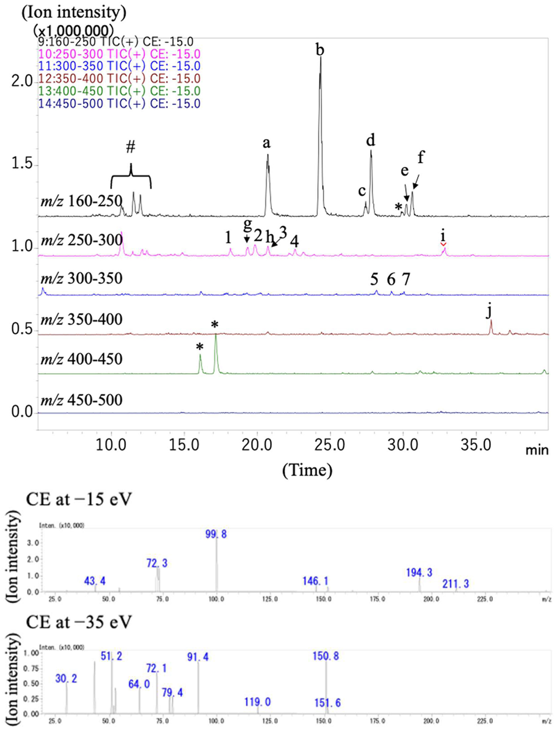 Click for large image | Figure 1. Mass chromatograms of 2-nitrophenylhydrazides of a cecal content of the rat in the HRP group in neutral loss scan mode (upper panel) and spectra of the product ions from precursor ions in peak 1 (lower panel). The peaks marked with lowercase in the chromatograms were estimated to be SCFAs and organic acids based on their retention time and m/z. The peaks marked with number were estimated to be modified amino acids consisting of SCFAs and amino acids on the basis of their m/z and mass spectra of product ion scanning. The peaks marked with sharp (#) were derived from the reagents and the peaks with asterisks could not be assigned to be any organic acids and modified amino acids. The product ion scan was carried out using collision energies at −35 and −15 eV. |
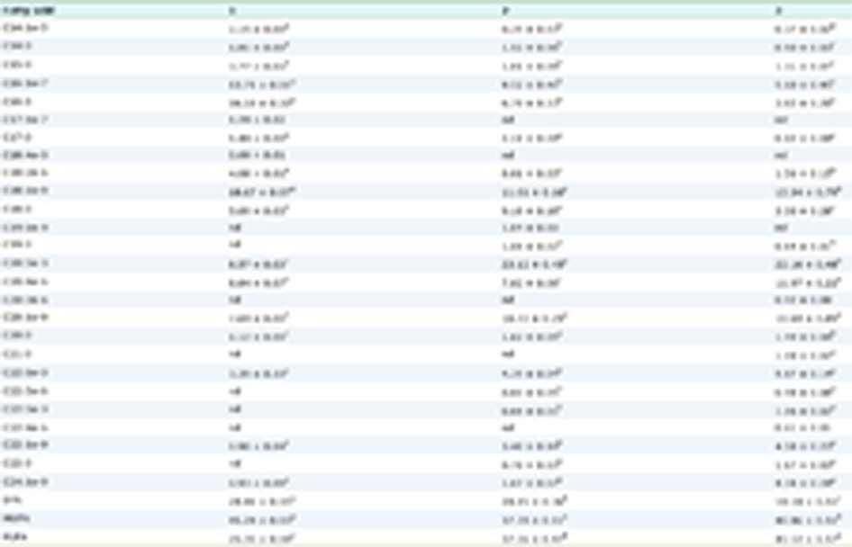 Click to view | Table 1. Summary of estimated SCFAs and organic acids in the rat cecal contents |
The precursor and product ions and the estimated structures of compounds in peaks 1–7 are summarized in Table 2. Subsequent MRM analysis using standards confirmed the presence of N-acetyl amino acids (NAAAs) listed in Table 2 in cecal contents. N-Propionyl-Ile/Leu, N-propionyl-Gly, and N-propionyl-Ala could not be validated due to unavailability of commercial standards. Peaks marked with asterisk (*) (Figure 1 upper panel) could not be assigned to known SCFAs, organic acids, or modified amino acids based on their MS/MS spectra. Peaks marked with # were derived from the derivatization reagents, as they were also detected in control injection of ultra-pure water processed with 2-NPH (data not shown).
Click to view | Table 2. Summary of estimated modified amino acids in the rat cecal contents |
3.2. Effects of RPH and pyroGlu-Leu on body weight gain and blood biochemical parameters
High-fat diet feeding did not significantly affect final body weight or total calorie intake (Table 3), indicating that the present 45% high-fat diet did not induce obesity. Plasma biochemical parameters, including AST, ALT, and creatinine, were not significantly altered by the high-fat diet, except for a significant increase in ALP, suggesting the absence of severe hepatic or renal dysfunction. In the PEL group, plasma TG levels were significantly higher than in the HF group; however, no other biochemical parameters differed significantly between RPH or PEL group and the HF group (Table 4).
Click to view | Table 3. Initial and final body weight and total food intake in rats |
Click to view | Table 4. Summary of blood biochemical values in rats |
3.3. Effects of RPH and pyroGlu-Leu on SCFAs in cecal content
Cecal SCFAs concentrations were quantified by LC-MS/MS in MRM mode. High-fat diet feeding significantly reduced the levels of acetic, propionic, and butyric acids (Figure 2a–c). Administration of RPH and pyroGlu-Leu did not significantly restore propionic or butyric acid levels compared to the HF group. Acetic acid levels were slightly but significantly increased in the HRP group (100 mg/kg body weight/day) compared to the HF group. No significant differences were observed among groups for other organic acids, including lactic, pyruvic, succinic, and malic acids (data not shown).
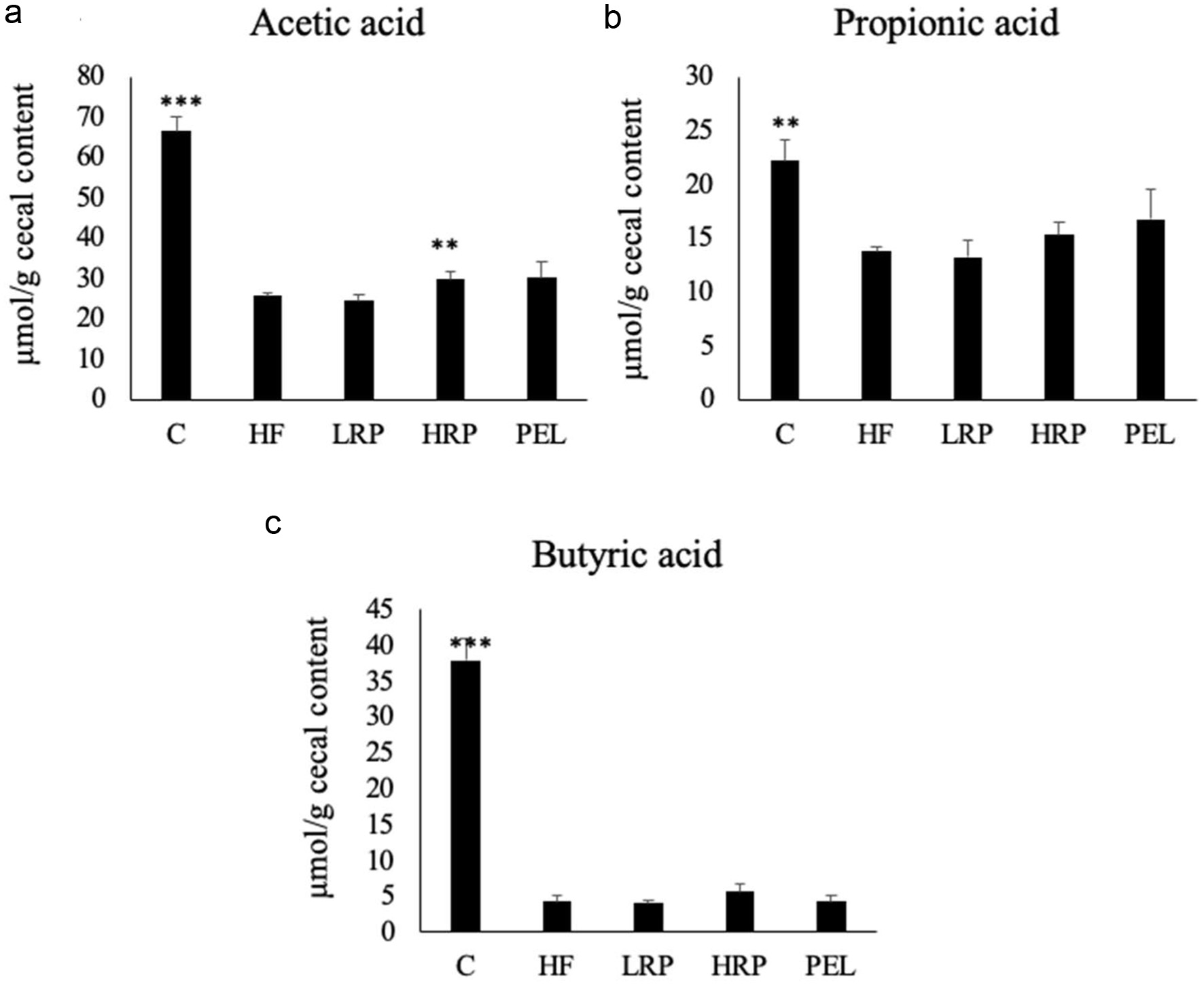 Click for large image | Figure 2. Concentrations of (a) acetic acid, (b) propionic acid, and (c) butyric acid in the cecal contents of rats. Data are shown as mean ± standard error (a–e, n = 4). Asterisks indicate significant differences compared to the HF group evaluated by Steel’s test (** p < 0.01, *** p < 0.001). |
3.4. Effects of RPH and pyroGlu-Leu on N-acetyl amino acids (NAAAs) in cecal contents
N-acetyl-Gly and N-acetyl-Ala were quantified by following derivatization with 2-NPH followed by LC-MS/MS analysis under the same chromatographic conditions used for their identification. Other NAAAs (N-acetyl-Ile, N-acetyl-Leu, and N-acetyl-Phe) were quantified without derivatization, as they were sufficiently retained under the present LC condition.
High-fat diet feeding significantly reduced the levels of all NAAA compared to C group (Figure 3). In contrast, administration of RPH at both doses and pyroGlu-Leu significantly restored the levels of N-acetyl-Gly, N-acetyl-Ala, and N-acetyl Ile (Figure 3a–c). N-Acetyl-Leu and N-Acetyl-Phe showed similar trends, although significant restoration was observed only in the low-dose RPH group (20 mg/kg/day) (Figure 3d, e).
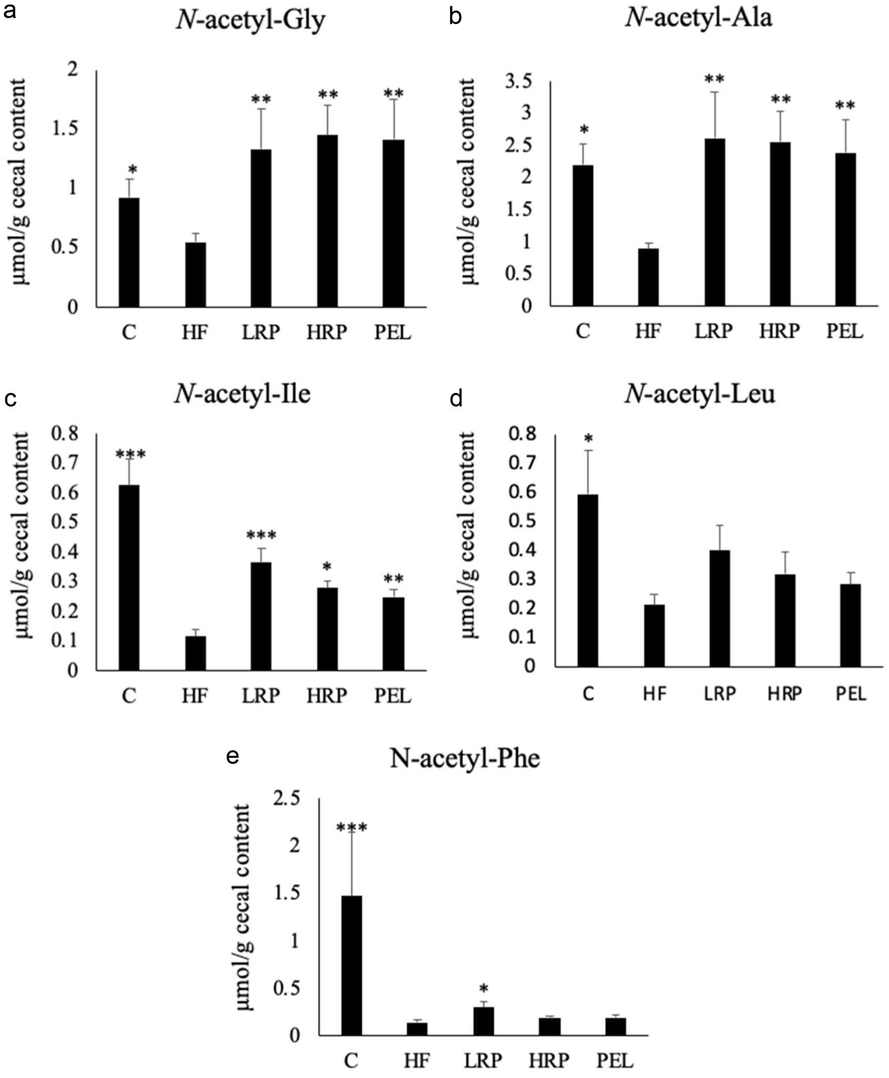 Click for large image | Figure 3. Concentrations of (a) N-acetyl-Gly, (b) N-acetyl-Ala, (c) N-acetyl-Ile, (d) N-acetyl-Leu, and (e) N-acetyl-Phe in the cecal content of rats. Data are shown as mean ± standard error (a–e, n = 4). Asterisks indicate significant differences compared to the HF group evaluated by Steel’s test (* p < 0.05, ** p < 0.01, *** p < 0.001). |
3.5. Effect of RPH and pyroGlu-Leu on fecal microbiota
Some fecal bacterial taxa could not be identified to species levels and are presented at family level. NMDS based on Bray-Curtis distances revealed no significant differences in overall microbiota composition among the HF, LRP, HRP, and PEL groups, as determined by pairwise Adonis test (Figure 4a).
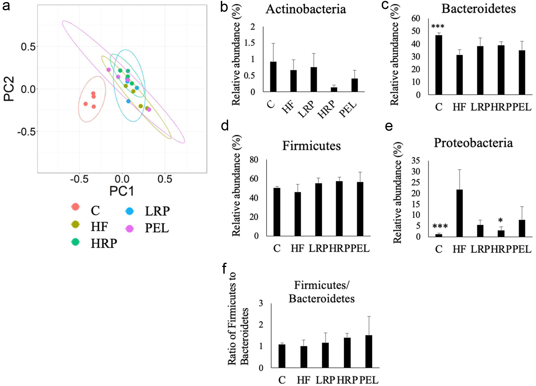 Click for large image | Figure 4. Non-metric multidimensional scaling (NMDS) plot of rat fecal microbiota based on Bray-Curtis distance (a) and the relative abundance of Actinobacteria (b), Bacteroidetes (c), Firmicutes (d), and Proteobacteria (e) and the ratio of Firmicutes to Bacteroidetes (f) in the rat fecal microbiota. Data are shown as mean ± standard error (b–f, n = 4). Asterisks indicate significant differences compared to the HF group by Steel test (* p < 0.05, *** p < 0.001). |
At the phylum level, the HF group exhibited a significantly lower relative abundance of Bacteroidetes and significantly higher levels of Proteobacteria compared with C group (Figure 4c and e). Administration of RPH at 100 mg/kg/day (the HRP group) significantly attenuated the high-fat-diet-induced increase in Proteobacteria (Figure 4e) with similar trends observed in the LRP and PEL groups. No significant differences were observed among groups in relative abundances of Actinobacteria and Firmicutes, nor in the Firmicutes/Bacteroidetes ratio (Figure 4b, d, and f).
At the taxon level, several differences in relative abundance were observed based on the Steel test. The HF group showed significantly lower abundances of Porphyromonadaceae (family level ID only) and significantly higher abundances of [Eubacterium] dolichum, Phascolarctobacterium faeccium, Enterococcus gallinarumcompared, and Clostridium symbiosum compared with C group (Figure 5b, g, k, l, and n). Administrations of RPH altered several taxa relative to the HF group. Low-dose RPH significantly increased Clostridiaceae (slash calls), [Eubacterium] dolichum, and Victivallaceae (family level ID only) (Figure 5a, b, and c), and decreased Arthrobacter creatinoliticus (Figure 5d). High-dose RPH significantly increased Bacteroides sp., Bacteroides thetaiotaomicron, Porphyromonadaceae (family level ID only), Eubacteriaceae (family level ID only), Blautia producta, Ruminococcus flavefaciens, and Phascolarctobacterium faecium (Figure 5e–k), while significantly decreasing Arthrobacter creatinoliticus, Enterococcus gallinarum, Lactobacillus animalis, and Clostridium symbiosum, (Figure 5d, l–n). However, none of these taxon-level differences remained statistically significant after Benjamini–Hochberg correction across the 130 taxa examined in each group comparison. Therefore, these findings were should be regarded as exploratory.
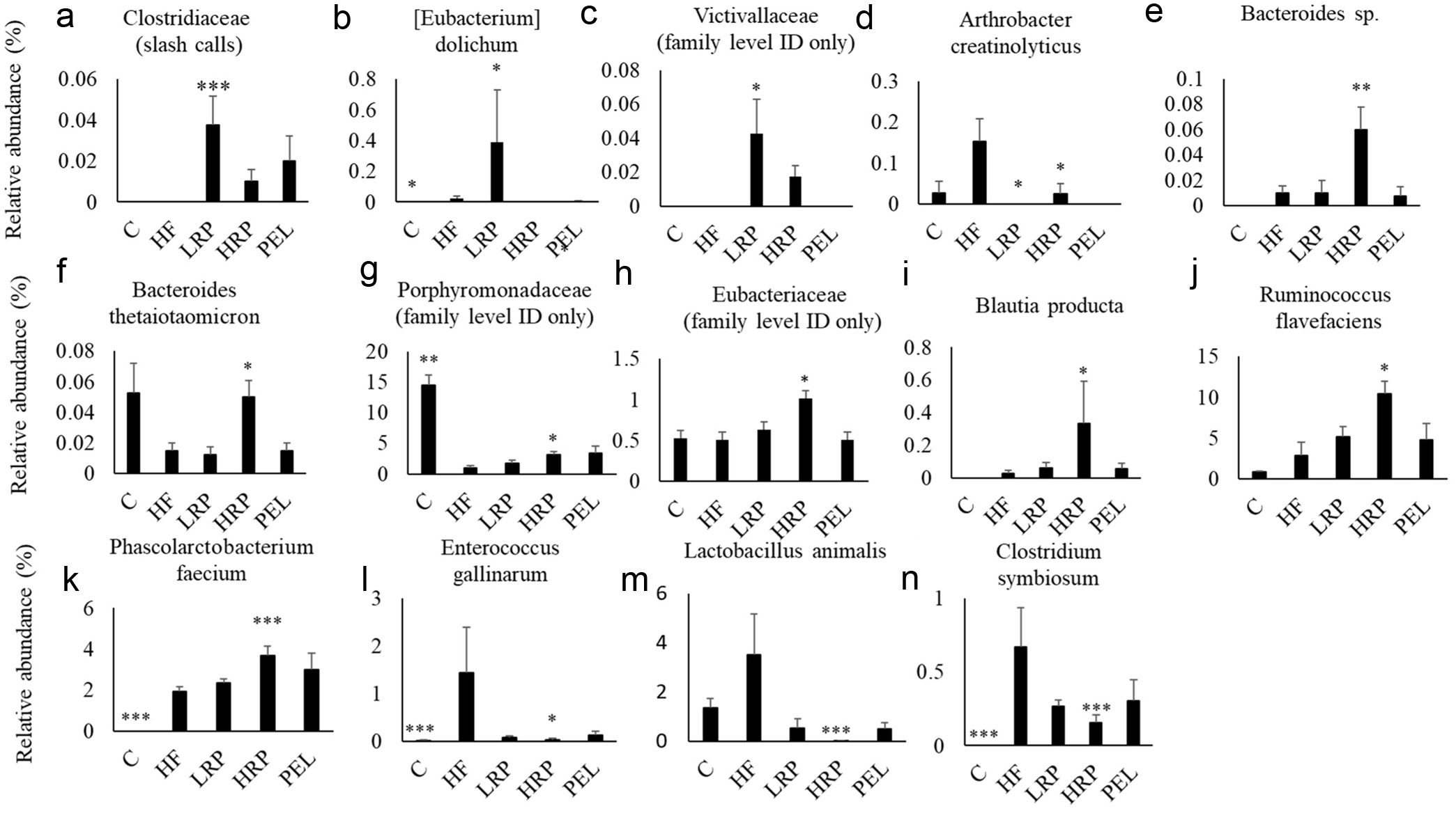 Click for large image | Figure 5. Relative abundance of Clostridiaceae (slash calls) (a), [Eubacterium] dolichum (b), Victivallaceae (family level ID only) (c), Arthrobacter creatinolyticus (d), Bacteroides sp. (e), Bacteroides thetaiotaomicron (f), Porphyromonadaceae (family level ID only) (g), Eubacteriaceae (family level ID only) (h), Blautia producta (i), Ruminococcus flavefaciens (j), Phascolarctobacterium faecium (k), Enterococcus gallinarum (l), Lactobacillus animalis (m), and Clostridium symbiosum (n) in the rat fecal microbiota. “Slash calls” indicate ambiguous assignments where sequences matched multiple taxa with similar confidence and could not be uniquely assigned to a single genus. Square brackets in [Eubacterium] dolichum denote a taxon that is currently considered misclassified in that genus and awaits formal taxonomic revision. “Family level ID only” indicates sequences that could be classified with sufficient confidence only to the family rank based on the 16S rRNA gene. Data are shown as mean ± standard error (A–N, n = 4). Asterisks indicate significant differences compared to the HF group by Steel’s test (* p < 0.05, *** p < 0.001). |
3.6. Correlation between fecal bacterial abundances and SCFAs and NAAAs
Spearman’s rank correlation coefficients between fecal bacterial species that differed significantly among groups and the levels of SCFAs and NAAAs are presented as a heat map (Figure 6). SCFAs and NAAAs exhibited similar directions of correlation (positive or negative) with the bacterial abundances; however, the strengths of these correlations differed. SCFAs showed strong positive correlations with Bacteroides thetaiotaomicron, Porphyromonadaceae (family level ID only), Ruminococcus flavefaciens, and Phascolarctobacterium faecium. NAAAs also correlated positively with these taxa but with weaker correlation strengths. The exact Spearman’s rank correlation coefficients (ρ) and their associated p-values underlying the heat map are provided in Table S3.
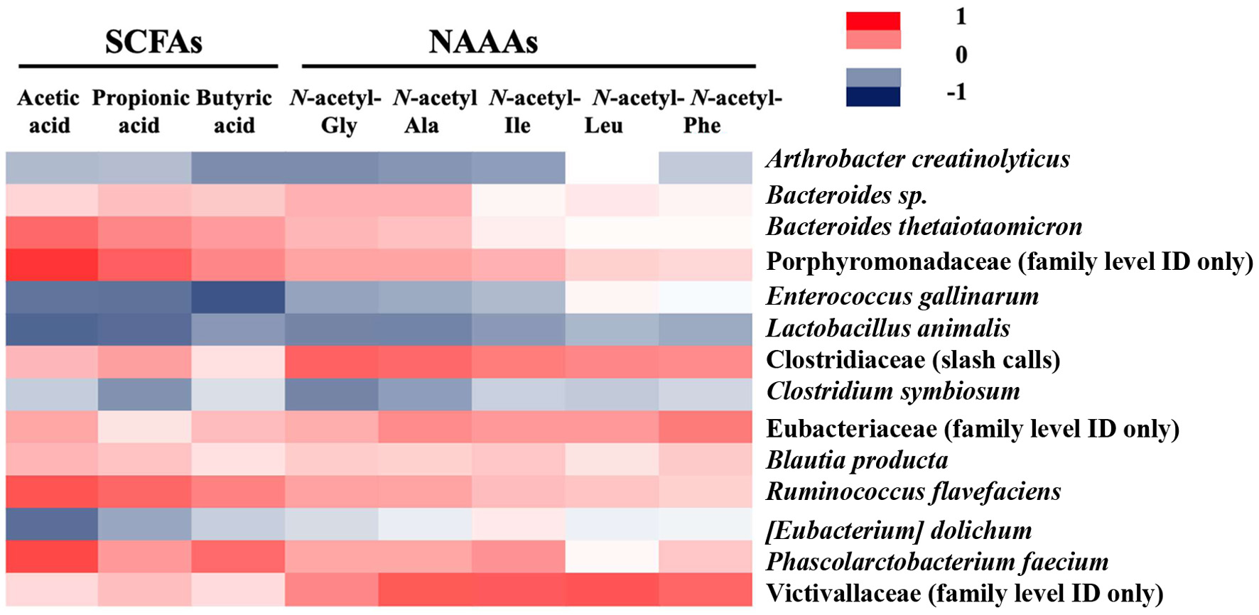 Click for large image | Figure 6. A heat map of Spearman’s rank correlation coefficients between fecal microbial species and SCFAs and NAAAs. |
In contrast, NAAAs exhibited stronger positive correlations than SCFAs with Clostridaceae (slash calls), Eubacteriaceae (family level ID only), and Victivallaceae (family level ID only). SCFAs showed stronger negative correlations with Enterococcus gallinarum and Lactobacillus animalis than did NAAAs. Additionally, acetic acid showed a particularly strong negative correlation with [Eubacterium] dolichum. Arthrobacter creatinolyticus exhibited negative correlations with several SCFAs and NAAAs.
| 4. Discussion | ▴Top |
Several studies have reported that short-chain pyroglutamyl peptides, such as pyroGlu-Leu and pyroGlu-Asn-Ile, exert beneficial effects on intestinal health, including amelioration of colitis in mouse models at low doses (0.1–1.0 mg/kg body weight) (Kiyono et al., 2016; Wada et al., 2013). These peptides have also been shown to modulate gut microbiota composition (Kiyono et al., 2016; Shirako et al., 2019; Wada et al., 2013). Shirako et al. (2019) demonstrated that pyroGlu-Leu improves high-fat-diet-induced dysbiosis by enhancing the secretion of rattusin, a host antimicrobial peptide produced in the small intestine. These studies used single synthetic pyroglutamyl peptides; however, pyroglutamyl peptides also naturally occur in fermented foods such as Japanese rice wine and miso, as well as in enzymatic hydrolysates of food proteins including wheat gluten and rice protein. Unlike fermented foods, enzymatic hydrolysates do not contain high levels of salt or alcohol, making them promising sources of pyroglutamyl peptides for use as functional food ingredients. The RPH used in the present study contained approximately 1.2 mg/g of pyroGlu-Leu and released an additional 1.2 mg/g after exopeptidase digestion (Miyauchi et al., 2022). Therefore, we compared the effects of RPH with those of synthetic pyroGlu-Leu on fecal microbiota and cecal metabolites in rats fed a high-fat diet. The doses of RPH used in this study (20 and 100 mg/kg body weight) corresponded to approximately 0.05 and 0.24 mg/kg body weight of pyroGlu-Leu, respectively, taking into account exopeptidase-mediated release.
High-fat-diet feeding is known to alter the gut microbiota, typically decreasing Bacteroidetes and increasing Firmicutes and Proteobacteria (Malesza et al., 2021; Murphy et al., 2015; Shirako et al., 2019) as well as reducing fecal SCFA concentrations (Brinkworth et al., 2009). SCFAs regulate inflammatory and immune responses in colonocytes and innate immune cells by acting as ligands of G-protein coupled receptors such as GPR41, GPR43, and GPR109A and by inhibiting histone deacetylases (Parada Venegas et al., 2019). These findings suggest that reductions in SCFAs due to high-fat diet-induced dysbiosis contribute to the onset and progression of diseases such as inflammatory bowel disease (IBD) and colorectal cancer. Indeed, high-fat-diet feeding accelerates the development of these diseases in animal models (Gruber et al., 2013; Lee et al., 2015; Shao et al., 2023). In addition to SCFAs, the present study identified N-acetyl amino acids (NAAAs) in cecal contents. Although several metabolomic studies have reported the presence of NAAAs in feces (Tian et al., 2024; Zierer et al., 2018), these compounds have not been quantified. Their producers and biosynthetic pathways remain unclear; however, their presence in fermented foods such as miso, soy sauce, and fish sauce (Nagao et al., 2024; Rahmadian et al., 2025) suggests that they are likely generated by microorganisms.
In the present study, feeding a 45% high-fat diet significantly altered the overall composition of the fecal microbiota (Figure 4a). However, unlike in our previous study using a 60% high-fat diet (Shirako et al., 2019), the Firmicutes/Bacteroidetes ratio was not significantly affected (Figure 4f). Furthermore, the present 5-week high-fat diet feeding did not increase body weight, in contrast to our previous study using the same animal strain and diet (Shirako et al., 2020). One plausible explanation for this difference is variation in the initial composition of the small-intestinal microbiota among the animals. Because metabolites produced by the small-intestinal microbiota contribute to high-fat-diet-induced obesity (Martinez-Guryn et al., 2018), the high-fat diet used in the present study may have induced only modest dysbiosis, possibly due to differences in the initial small-intestinal microbiota of the rats.
Despite the absence of obesity or liver damage, the high-fat diet significantly decreased cecal SCFA levels (Figure 2). Administration of RPH or pyroGlu-Leu did not restore SCFA levels or reverse the overall microbiota composition altered by the high-fat diet. However, both doses of RPH and pyroGlu-Leu significantly restored the levels of N-acetyl-Gly, N-acetyl-Ala, and N-acetyl-Ile (Figure 3), and altered the relative abundances of several bacterial taxa at the phylum and species levels (Figures 4 and 5). Recently, Kvitne et al. (2025) reported that children with very early-onset IBD exhibit lower abundances of SCFA-producing bacteria and reduced fecal levels of NAAAs such as N-acetyl-Met and N-acetyl-Ile/Leu, with positive correlations between these bacteria and NAAAs. These findings suggest that NAAAs may be synthesized by SCFA-producing bacteria and may serve as biomarkers reflecting their abundance or metabolic activity. Consistent with this, the present study showed that SCFAs and NAAAs correlated in the same direction with several bacterial species; however, the strengths of these correlations differed (Figure 6). Notably, the abundances of Clostridaceae, Eubacteriaceae, and Victivallaceae showed stronger positive correlations with NAAAs than with SCFAs. These families include known SCFA-producing bacteria (Esquivel-Elizondo et al., 2017; Mukherjee et al., 2020; Zoetendal et al., 2003). These results suggest that oral administration of RPH and pyroGlu-Leu may enhance the growth or metabolic activity of SCFA-producing bacteria in a manner that increases NAAA production without necessarily increasing SCFA levels.
The mechanisms underlying these observations remain unclear. One possibility is that NAAAs respond more rapidly or sensitively to changes in SCFA-producing bacteria than SCFAs, which often require larger shifts in microbial fermentation to change measurably. Alternatively, RPH and pyroGlu-Leu may preferentially promote bacteria capable of producing N-acetyl amino acids or modulate metabolic pathways within SCFA-producing bacteria, leading to increased NAAA synthesis. Further studies are needed to clarify these mechanisms, but the present findings indicate that NAAAs may serve as early and sensitive indicators of microbiota-host co-metabolism.
The biological roles of NAAAs in the host remain largely unknown. In this study, administration of RPH and pyroGlu-Leu restored NAAA levels before the onset of adverse effects typically associated with high-fat diet feeding. Thus, the recovery of NAAAs may contribute to the beneficial effects of pyroglutamyl peptides on intestinal health reported previously (Kiyono et al., 2016; Wada et al., 2013). Takakuwa et al. (2019) reported that SCFAs and amino acids such as butyrate and leucine stimulate α-defensin secretion from mouse Paneth cells. Therefore, NAAAs restored by RPH and pyroGlu-Leu administration may modulate innate immunity in the intestine, similarly to pyroGlu-Leu, amino acids, and SCFAs. Further studies on the biological activities of N-acetyl amino acids are currently underway.
This study has several limitations. First, the sequencing analysis was outsourced, and detailed quality-control metrics—including sequencing depth, read-quality summaries, filtering criteria, chimera removal, and taxonomic-assignment thresholds—together with the raw sequence files were part of the contract laboratory’s internal workflow and could not be retrieved retrospectively. Absolute bacterial quantification and compositional differential-abundance analysis were also not performed; therefore, the microbial data were analyzed only as relative abundances, and the taxon-level findings should be regarded as exploratory. Second, the baseline composition of the gut microbiota was not assessed, so comparability among groups before the intervention could not be directly confirmed, and the proposed contribution of the initial small-intestinal microbiota to the absence of body-weight gain remains speculative. Third, only endpoint microbiota profiles were examined, and the relatively small sample size (n = 4 per group) may limit the statistical power to detect subtle differences. These limitations should be addressed in future studies incorporating baseline and longitudinal microbiota profiling, absolute quantification, and larger group sizes.
Furthermore, modulation of the gut microbiota and its metabolites by RPH or pyroGlu-Leu may have the potential to support human health by contributing to the maintenance of a favorable intestinal environment. Based on body-surface-area scaling, the RPH doses used in this study correspond to estimated human-equivalent intakes of approximately 0.19–0.97 g/day for a 60-kg adult, whereas the pure pyroGlu-Leu dose corresponds to approximately 9.7 mg/day. The estimated RPH intakes would provide or potentially release approximately 0.47–2.34 mg of pyroGlu-Leu per day. Although pyroglutamyl peptides are absorbed by small-intestinal tissue, our previous study (Miyauchi et al., 2022) showed that they only small parts of them enter the bloodstream. Therefore, body-surface-area scaling may not fully reflect their physiological behavior. Nevertheless, even when considering intake per body weight, the estimated human-equivalent doses of RPH and pyroGlu-Leu remain realistic. Human intervention studies are required to determine whether comparable effects occur in humans and to establish effective intake levels.
| 5. Conclusion | ▴Top |
The present study demonstrated the presence of NAAAs in the rat cecal contents by neutral loss scanning using LC-MS/MS following 2-NPH derivatization. Interestingly, the high-fat diet used in the present study significantly decreased cecal concentrations of NAAAs and SCFAs in rats through changes in fecal microbiota. However, administration of RPH and pyroGlu-Leu restored the levels of NAAA but not those of SCFA. Cecal concentrations of NAAA and SCFA showed similar positive correlations with the abundances of several SCFA-producing bacteria; however, the strength of these correlations differed between SCFAs and NAAAs. Collectively, these findings raise the possibility that NAAA represent an early metabolic signature of beneficial adaptations in SCFA-producing bacteria induced by pyroglutamyl peptides.
| Supplementary material | ▴Top |
Table S1. Daily water intake (mL) of rats during the 5-week experimental period.
Table S2. Actual daily doses (mg/kg body weight/day) of RPH and pyroGlu-Leu administered to rats during the 5-week experimental period.
Table S3. Spearman’s rank correlations between cecal metabolite levels and fecal bacterial relative abundances.
Acknowledgments
We would like to thank to the Kyoto Louis Pasteur Center for Medical Research for allowing us to perform animal experiments in their facility.
| References | ▴Top |