Figures
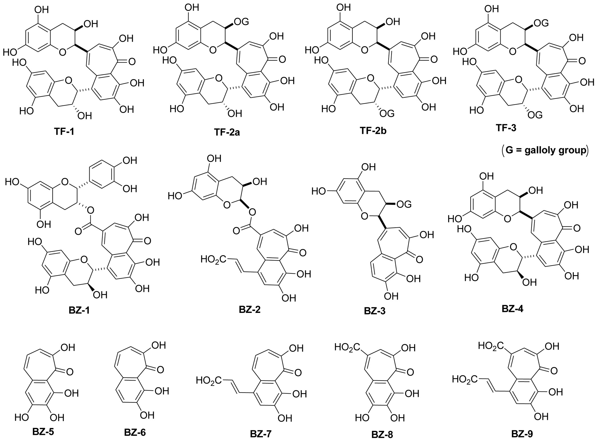
Figure 1. Structure of benzotropolone derivatives. Structures of the nine different benzotropolone derivatives (BZ-1 to BZ-9) are illustrated. For comparison, the four theaflavin isomers such as theaflavin (TF-1), theaflavin-3-O-gallate (TF-2), theaflavin-3′-O-gallate (TF-2′) and theaflavin-3,3′-O-O-digallate (TF-3) with the benzotropolone moiety as their core structure are also shown.
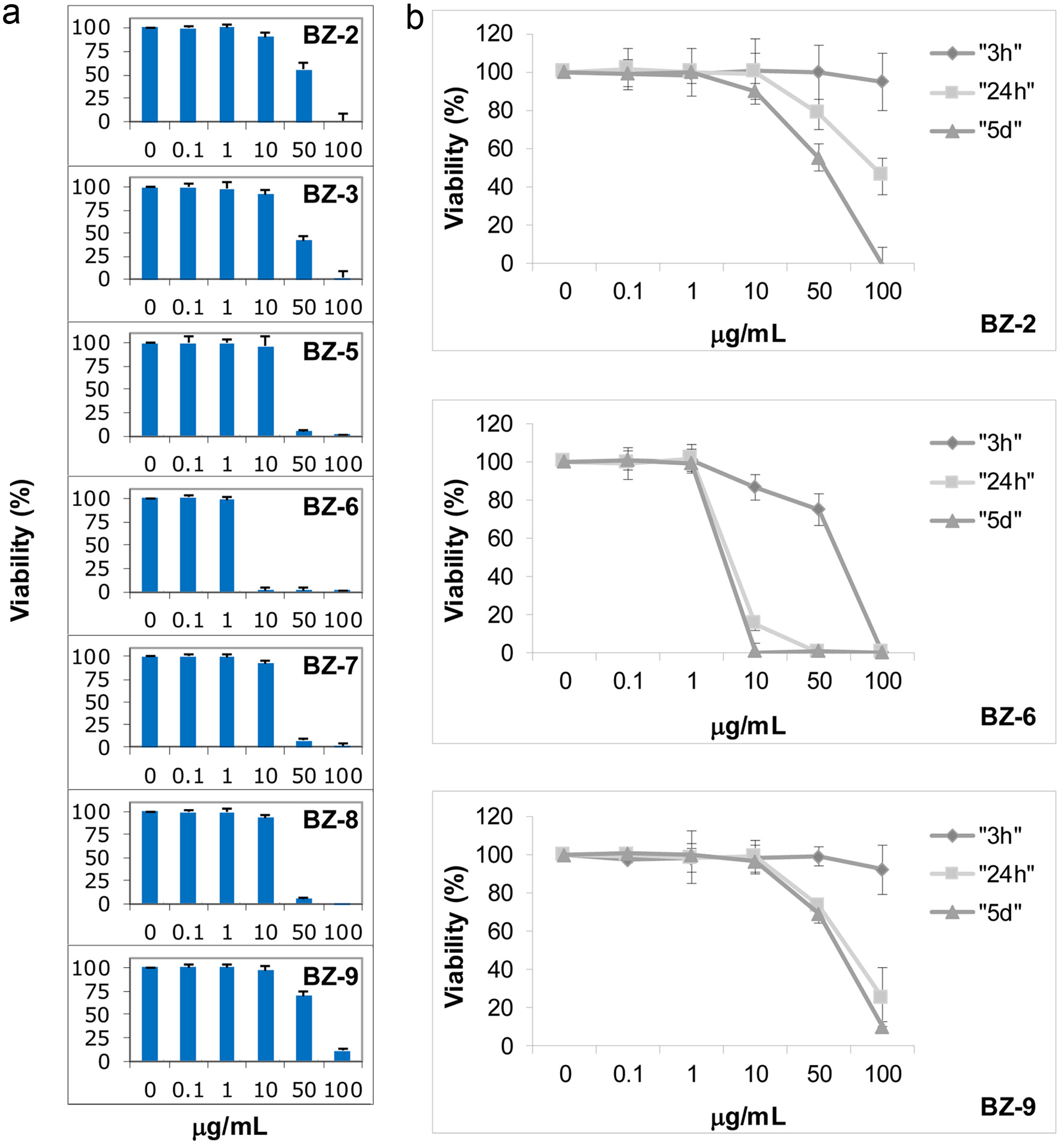
Figure 2. Effects of benzotropolone derivatives on cell viability. Human monocytes (U-937) were treated with different BZ derivatives at indicated concentrations and measured by the MTT assay. Mean values ± standard deviation of at least three independent experiments is shown. Cells were exposed to BZs either at different concentrations for 5 days (a) or different concentrations of BZ-2, BZ-6 and BZ-9 were incubated for 3 h, 24 h, or 5 days as indicated by different symbols (rhombus, square, and triangle), respectively (b).
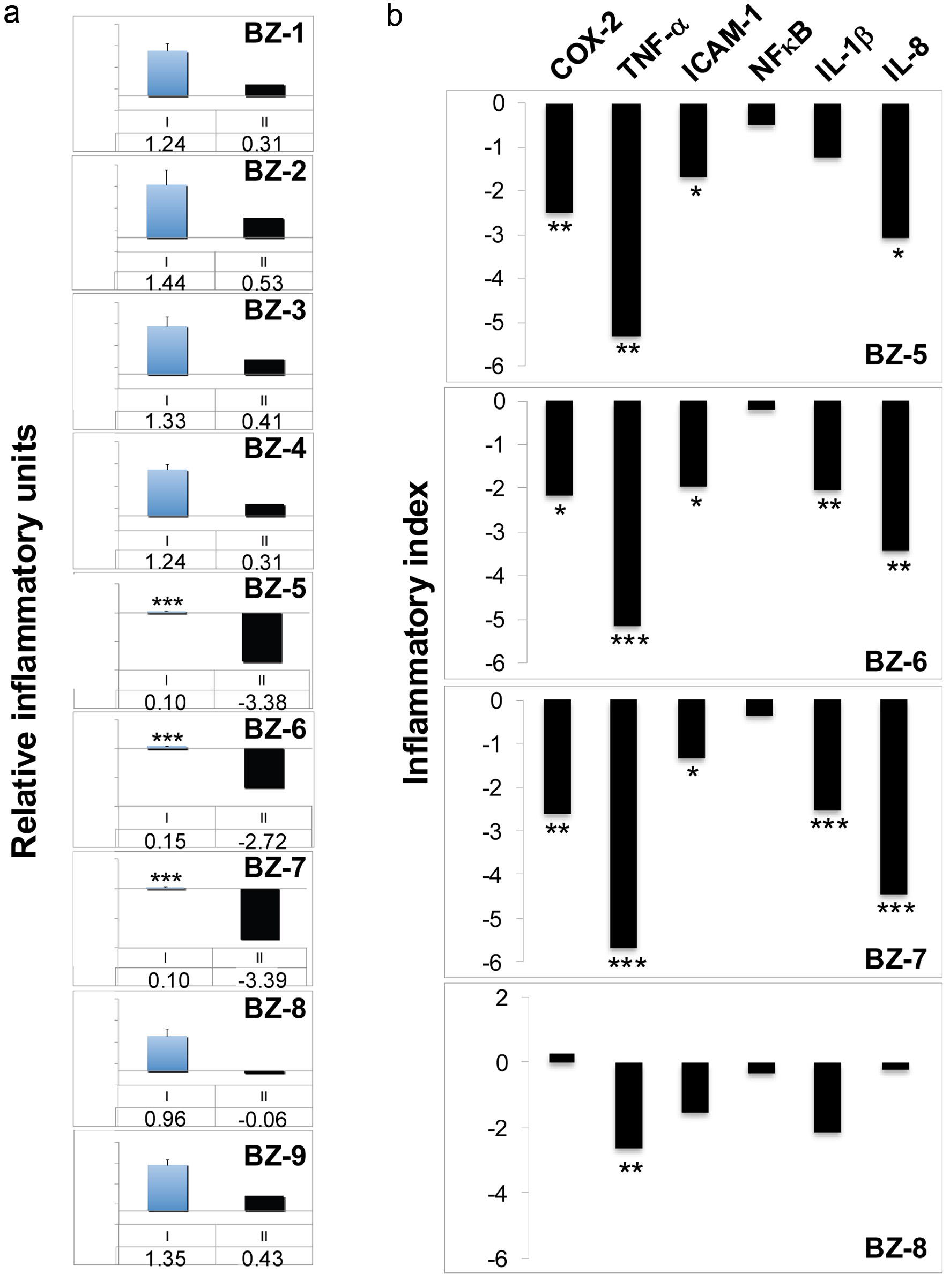
Figure 3. Effects of benzotropolone derivatives on the expression of inflammatory genes. Human monocytes (U-937) were treated with TPA (20nM) either alone or in combination with different BZs (50 µg/ml) for 3h. After RNA isolation and reverse transcription, expression of either COX-2 (a) or (b), a panel of different inflammatory genes (COX-2, TNF-α, ICAM-1, NFκB, IL-1β, and IL-8) was analyzed by TaqMan qPCR analysis. The expression of glyceraldehyde-3-phosphate dehydrogenase (GAPDH) was used as internal control. After normalization to GAPDH according to the delta-delta CT method, gene expression is expressed as a ratio of the mean experimental channel (TPA + BZ) to the mean control channel (TPA alone). Ratios converted into Log2 values are expressed as inflammatory index as described in Materials and Methods. *, **, and *** indicate significant differences from the control group with P < 0.05, 0.01 or 0.001, respectively, as analyzed by the student’s t-test of at least three independent experiments. (a) COX-2 expression is either shown as mean ratios + standard deviation indicated by blue columns (I, mean value below) or as inflammatory indices indicated by black columns (II, inflammatory index below). (b) Inflammatory indices of COX-2, TNF-α, ICAM-1, NFκB, IL-1β, and IL-8 are shown for BZ-5, BZ-6, BZ-7 and BZ-8 indicated by black columns.
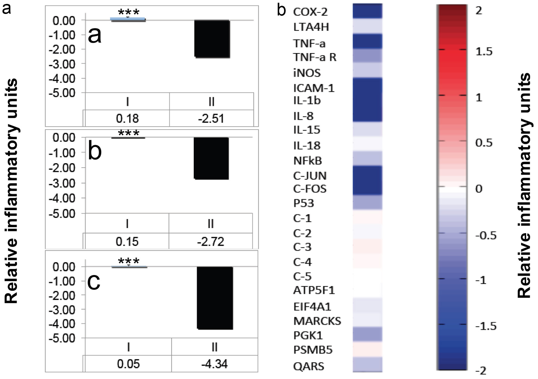
Figure 4. Effects of BZ-6 in U-937 cells. (a) Human monocytes (U-937) were treated with TPA (20nM) either alone or in combination with different concentrations of BZ-6 (25, 50 and 100 µg/ml) for 3h as indicated (a, b or c, respectively). After RNA isolation and reverse transcription, expression of COX-2 was analyzed by TaqMan qPCR analysis. The expression of glyceraldehyde-3-phosphate dehydrogenase (GAPDH) was used as internal control. After normalization to GAPDH, gene expression was calculated as ratios (I) or inflammatory indices (II), respectively. *** indicate significant differences from the control group with P < 0.001 as analyzed by the student’s t-test of at least three independent experiments. (b) U-937 cells were treated with TPA (20nM) either alone or in combination with BZ-6 (50 µg/ml) for 3h. After RNA isolation and reverse transcription, expression of COX-2, LTA4 hydrolase, TNF-α, TNF-α receptor, iNOS, ICAM-1, IL-1β, IL-8, IL-15, IL-18, NFκB, c-Jun, c-Fos, and p53 was analyzed by a proprietary Oligo microarray as described in Material and Methods. Artificial DNA sequences were used as control genes (C-1 - C-5) for normalization. As housekeeping genes, ATP5F1, EIF4A1, MARCKS, PGK1, PSMB5, and QARS were employed. Gene expression is shown as relative inflammatory units by cluster analysis. Upregulation is indicated by red colors, downregulation by blue colors, whereas no regulation is indicated by white colors. One set of representative data is shown of two independent experiments.
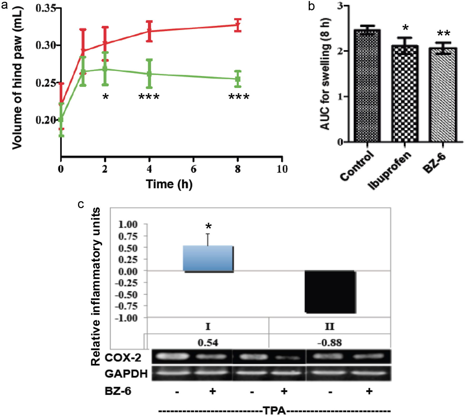
Figure 5. Effects of BZ-6 on carrageenan-induced paw edema in mice and in Caco-2 cells. (a, b) Mice received a single oral dose of control vehicle (tocopherol-stripped corn-oil), BZ-6 (250 mg/kg), or ibuprofen (100 mg/kg), respectively. 1 h after treatment, all groups received an injection of 0.1 mL of carrageenan (1% in water) into the plantar side of the right hind paw (n = 6 for each group per time point). The mice were then euthanized 1, 2, 4, and 8 h after carrageenan injection. The degree of swelling was determined by paw volume displacement measured at indicated times by a digital hydroplethysmometer. Results are expressed as mean ± SD of the volume of hind paw (mL) for different time points (a) or as area under the curve (AUC) units after 8 h (b), respectively. (c) Human colorectal carcinoma cells (Caco-2) were treated with TPA (20nM) either alone or in combination with BZ-6 (50 µg/ml) for 3h. After RNA isolation and reverse transcription, gene expression of COX-2 and GAPDH was analyzed by RT-PCR and quantified using densitometry. COX-2 expression normalized to GAPDH is expressed either as ratio of the mean experimental channel (TPA + BZ) to the mean control channel (TPA alone) ± standard deviation (I) or inflammatory index (II), respectively. Mean values + standard deviation of three independent experiments with representative blots are shown in the histograms. *, **, and *** indicate significant differences from the control group with P < 0.05, 0.01 or 0.001, respectively as analyzed by ANOVA (a, b) or students t-test (c).