Figures
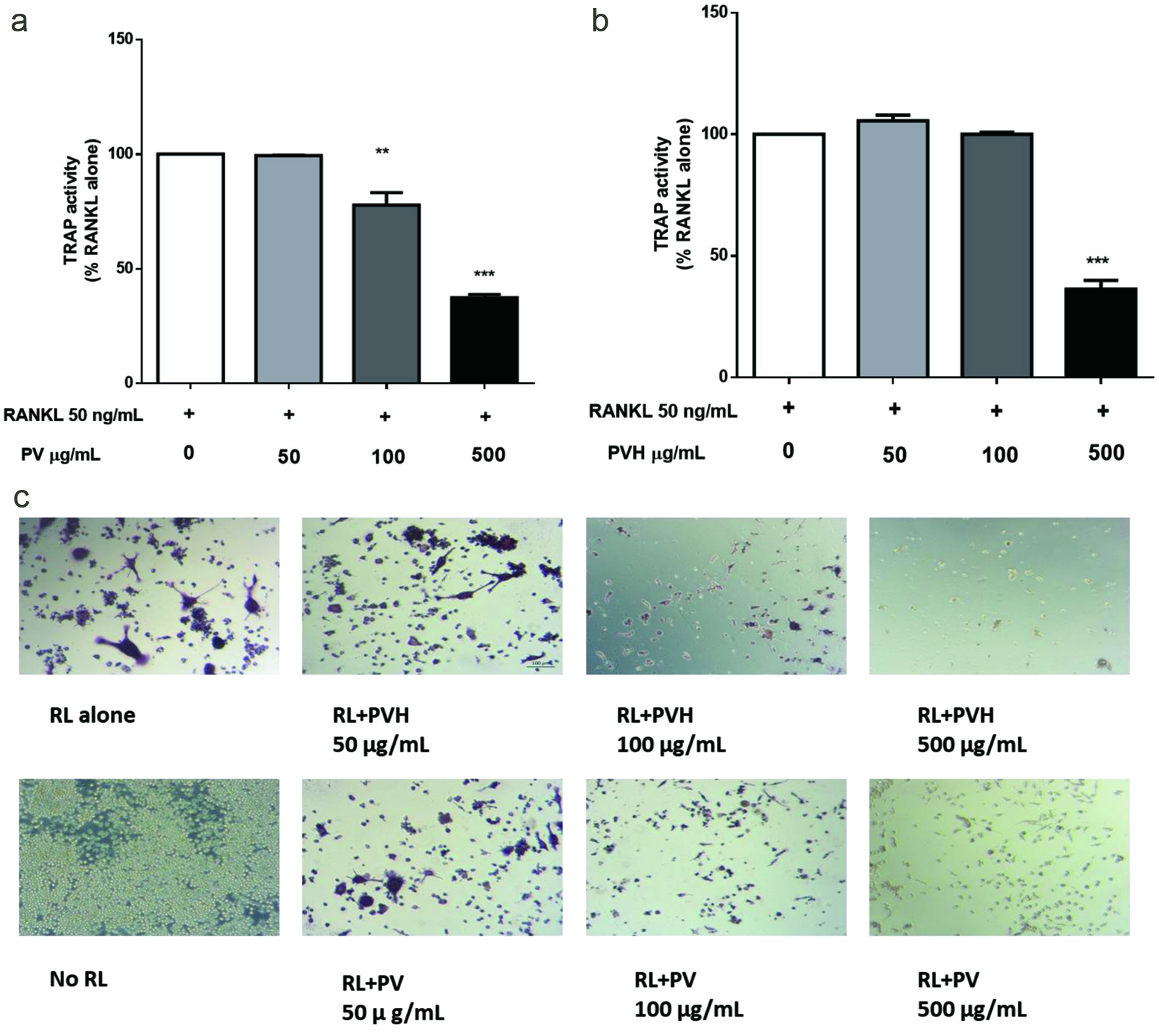
Figure 1. Effects of PV/PVH on the formation of TRAP-positive cells in RANKL-stimulated RAW264.7 cells. RAW264.7 cells were incubated with 50 ng/ml RANKL for 24 hr followed by PV or PVH for another 5 days, then the cells were lysed by 0.2% Triton X-100. TRAP activity in cell lysate was determined by using Sigma’s TRAP kit (CS0740). (a) TRAP activity of PV treated RAW264.7 cells; (b) TRAP activity of PVH treated RAW264.7 cells, (c) A set of representative images of cells stained with Sigma’s TRAP staining kit (387A-1KT). Results were expressed as the mean ± standard error of the mean (n = 3). *, ** and *** indicate p < 0.05, p < 0.01 and p < 0.001 respectively, as compared to the group of RANKL alone. (RL, RANKL; PV, phosvitin; PVH, phosvitin hydrolysate)
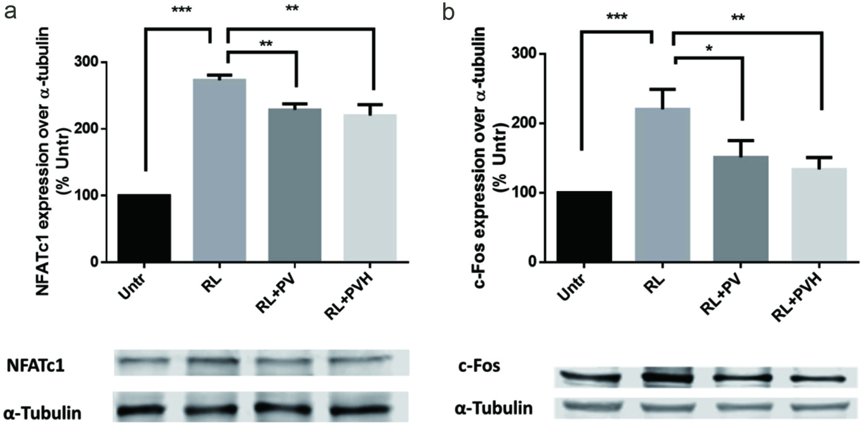
Figure 2. Effects of PV/PVH on osteoclastogenesis transcription factors in RANKL-stimulated RAW264.7 cells. RAW264.7 cells were cultured with RANKL (50 ng/ml) for 24 hr and followed by PV (500 µg) or PVH (500 µg) for another 72 hr. A: Whole cell lysates were analyzed by Western blot using antibodies against NFATc1. A set of representative images was shown. B: Whole cell lysates were analyzed by Western blot using antibodies to c-Fos. A set of representative images was shown. Results were expressed as the mean ± standard error of the mean (n = 3). *, ** and *** indicate p < 0.05, p < 0.01 and p < 0.001 respectively, as compared to the group of RANKL alone. (Untr, untreated group without RANKL or PV/PVH; RL, RANKL; PV, phosvitin; PVH, phosvitin hydrolysate)
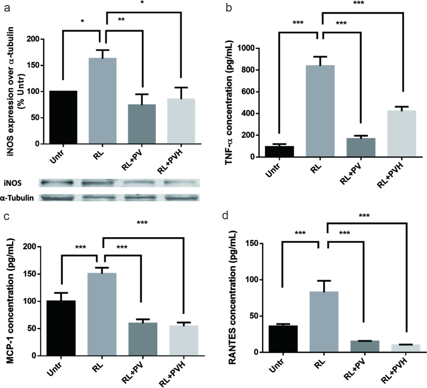
Figure 3. Effects of PV/PVH on inflammation protein expression/secretion in RANKL-stimulated RAW264.7 cells. RAW264.7 cells were cultured for 24 hr. Then, cells were incubated with either PV (500 µg) or PVH (500 µg) for 24 hr. RANKL (50 ng/ml) was added to stimulate inflammatory cytokines/chemokines secretion for 16 hr. At the end of culture, medium was collected and analyzed by using corresponding ELISA kits or the cell lysates were analyzed by Western blot. (a) Whole cell lysates were analyzed by Western blot using antibodies against iNOS. A set of representative images was shown. (b) TNF-α secretion in cell medium. (c) MCP-1 secretion in cell medium. (d) RANTES secretion in cell medium. Results were expressed as the mean ± standard error of the mean (n = 3). *, ** and *** indicate p < 0.05, p < 0.01 and p < 0.001 respectively, as compared to the group of RANKL alone. (Untr, untreated group without RANKL or PV/PVH; RL, RANKL; PV, phosvitin; PVH, phosvitin hydrolysate)
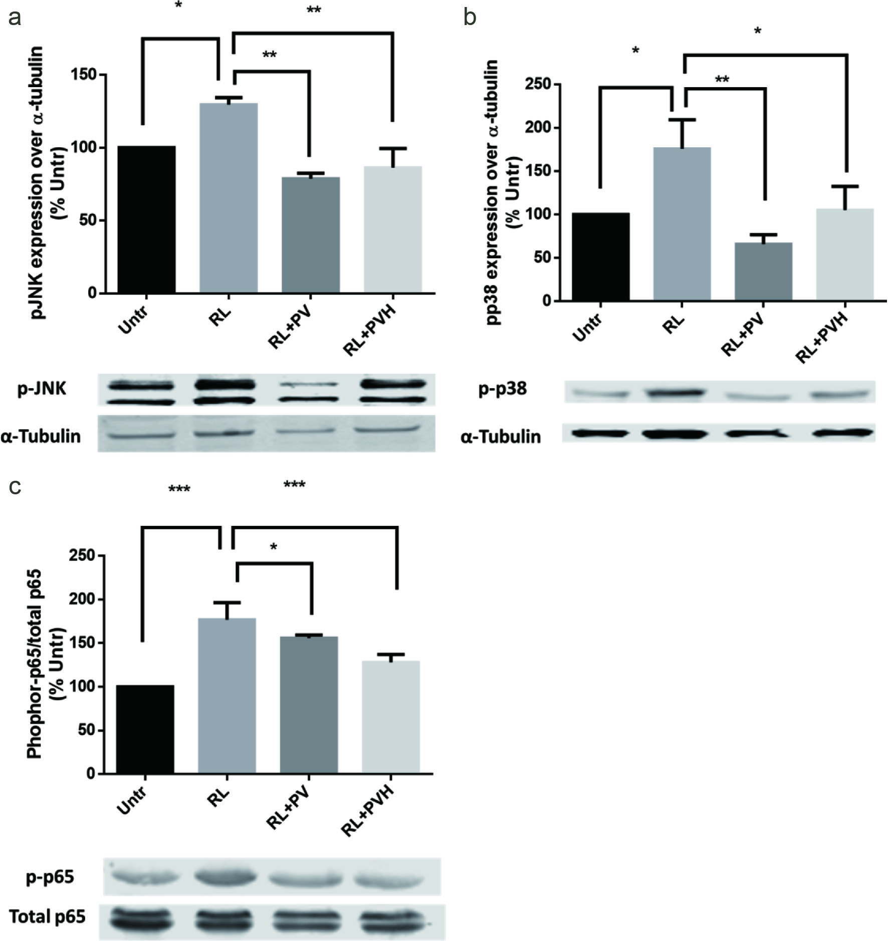
Figure 4. Effects of PV/PVH on different signaling pathways in RANKL-stimulated RAW264.7 cells. RAW264.7 cells were incubated with either PV (500 µg) or PVH (500 µg) for 48 hr, followed by treatment with RANKL (50 ng/ml) for 30 min for JNK/P36, or 45 min for P-65 expression. (a) Whole cell lysates were analyzed by Western blot using antibodies against JNK and phosphor-JNK. A set of representative images was shown. (c) Whole cell lysates were analyzed by Western blot using antibodies against p38 and phosphor-p38. A set of representative images was shown. (a) Whole cell lysates were analyzed by Western blot using antibodies against P65 and phosphor-P65. A set of representative images was shown. Results were expressed as the mean ± standard error of the mean (n = 3). *, ** and *** indicate p < 0.05, p < 0.01 and p < 0.001 respectively as compared to the group of RANKL alone. (Untr, untreated group without RANKL or PV/PVH; RL, RANKL; PV, phosvitin; PVH, phosvitin hydrolysate)