Figures
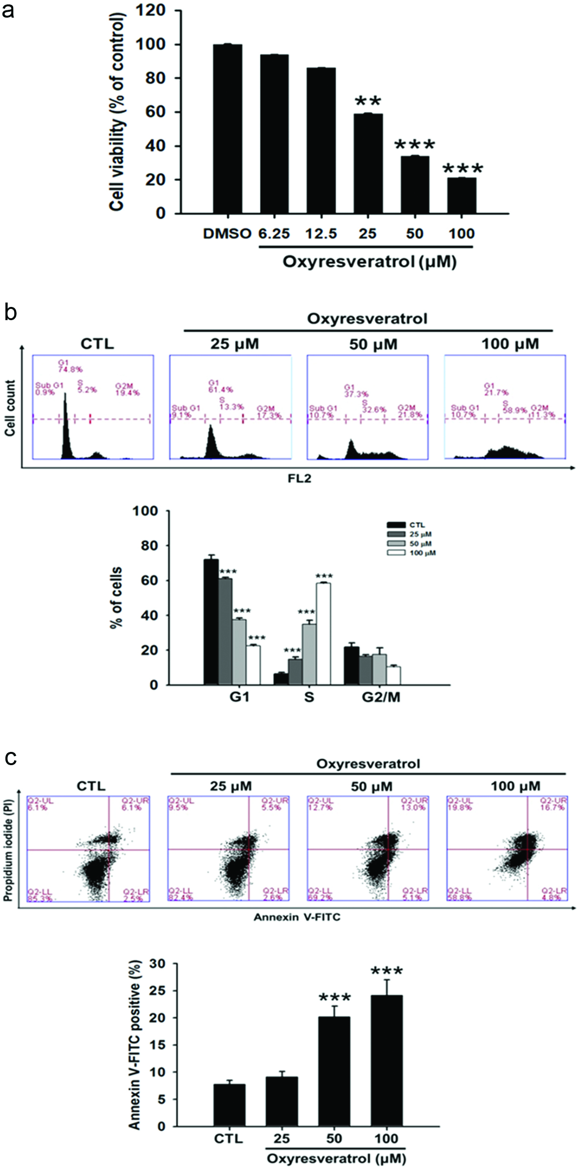
Figure 1. Effects of oxyresveratrol on (a) cytotoxicity, (b) cell cycle progression, and (C) apoptosis of NCI-H520 cells. NCI-H520 cells were treated with oxyresveratrol for 24 h. The cytotoxicity was analyzed by an MTT assay, and cell cycle distributions were then analyzed by flow cytometry. Apoptosis in NCI-H520 cells was examined by Annexin V-FITC/propidium iodide (PI) binding and analyzed by flow cytometry. Data are presented as the mean ± SD of three experiments each conducted in triplicate. **p < 0.01 and ***p < 0.001 compared to the DMSO-treated group.
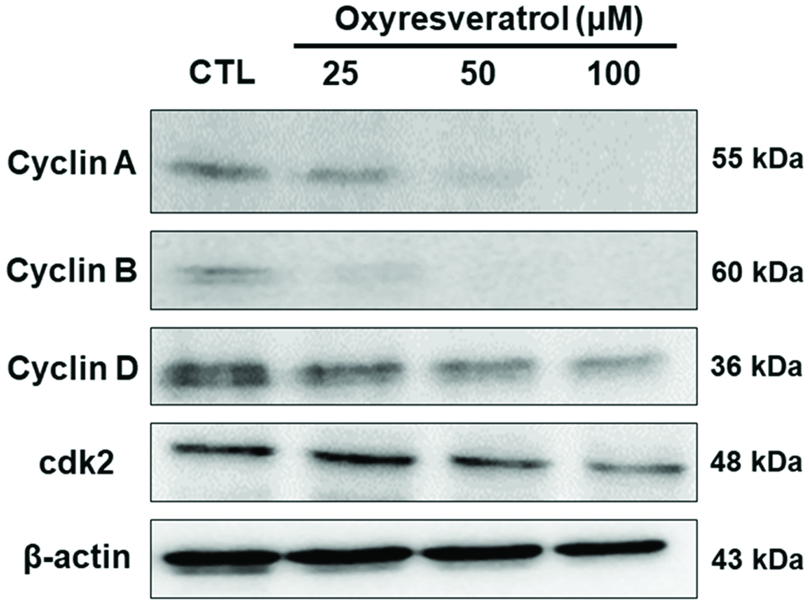
Figure 2. Effects of oxyresveratrol on cell-cycle-related proteins in NCI-H520 cells. NCI-H520 cells were treated with 25, 50, or 100 μM oxyresveratrol for 24 h, and then whole-cell lysates were prepared and subjected to Western blotting using antibodies against the indicated proteins. β-Actin was used as a loading control. Bands were analyzed by ImageJ software (National Institutes of Health) and normalized to β-actin.
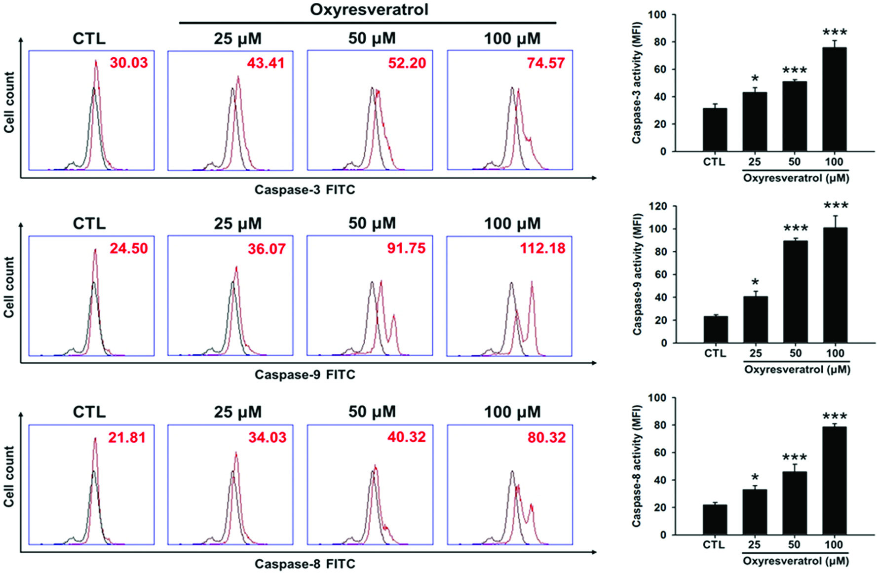
Figure 3. Effects of oxyresveratrol on caspase-3, -8, and -9 activities in NCI-H520 cells. NCI-H520 cells were treated with 25, 50, or 100 μM oxyresveratrol for 24 h and then the activities of caspases-3, -8, and -9 were determined by flow cytometry. Black line: mock control of DMSO-treated cell control; red line: cells treated with oxyresveratrol. Data are presented as the mean ± SD of three experiments each conducted in triplicate. *p < 0.05, **p < 0.01, and ***p < 0.001 compared to the DMSO-treated group.
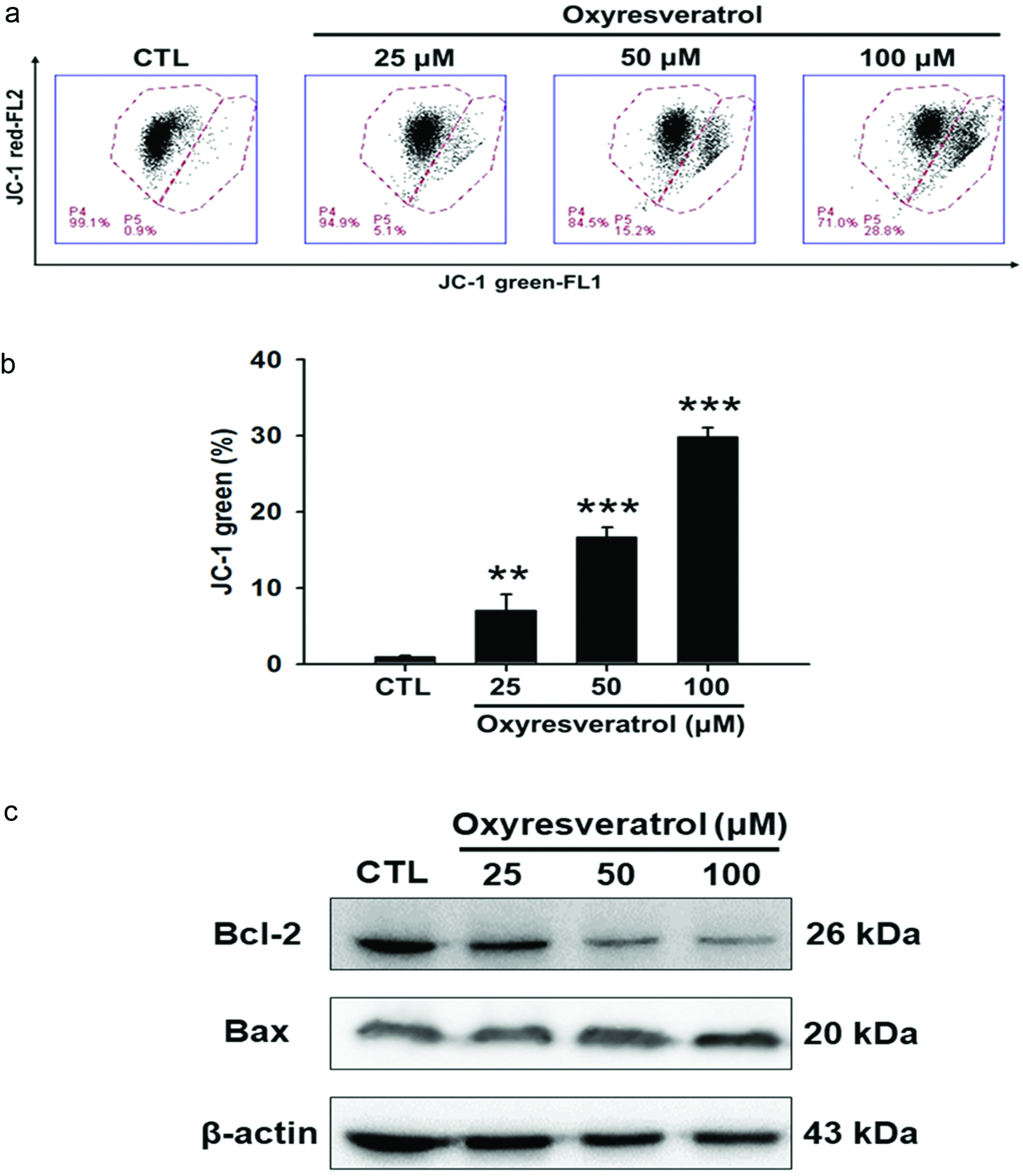
Figure 4. Effect of oxyresveratrol on the mitochondrial pathway of apoptosis in NCI-H520 cells. NCI-H520 cells were treated with 25, 50, or 100 μM oxyresveratrol for 24 h. (a) Disruption of the mitochondrial membrane potential. Data are presented as the mean ± SD of three experiments each conducted in triplicate. **p < 0.01 and ***p < 0.001 compared to the DMSO-treated group. (b) Bcl-2 and Bax were examined by a protein gel blot analysis. β-Actin was used as a loading control. Bands were analyzed by ImageJ software (National Institutes of Health) and normalized to β-actin.
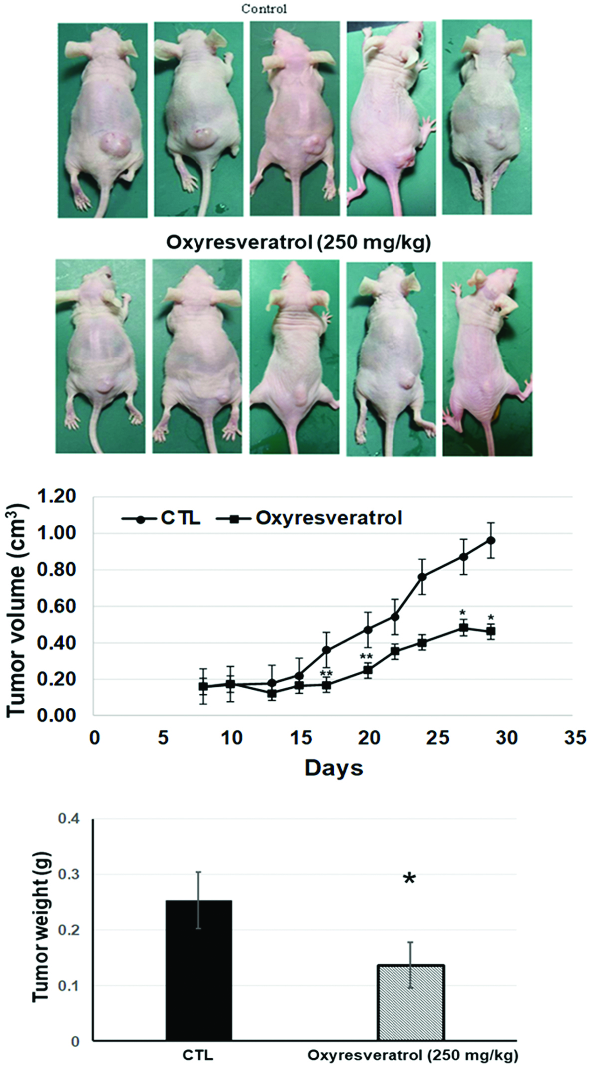
Figure 5. Oxyresveratrol inhibited NCI-H520 tumor growth in nude mice. NCI-H520 cells (107) were subcutaneously injected into mice and allowed to grow up to approximately 10 mm3 in size. Oxyresveratrol (250 mg/kg) and a vehicle control were administered intraperitoneally daily for three weeks until day 30 (n = 5). Photograph of tumor tissues (a), the time course of tumor sizes (b), and the tumor weight on day 30. Quantified data are presented as the mean ± SD (n = 5). *p < 0.05 and **p < 0.01 of comparisons between the vehicle and oxyresveratrol groups.