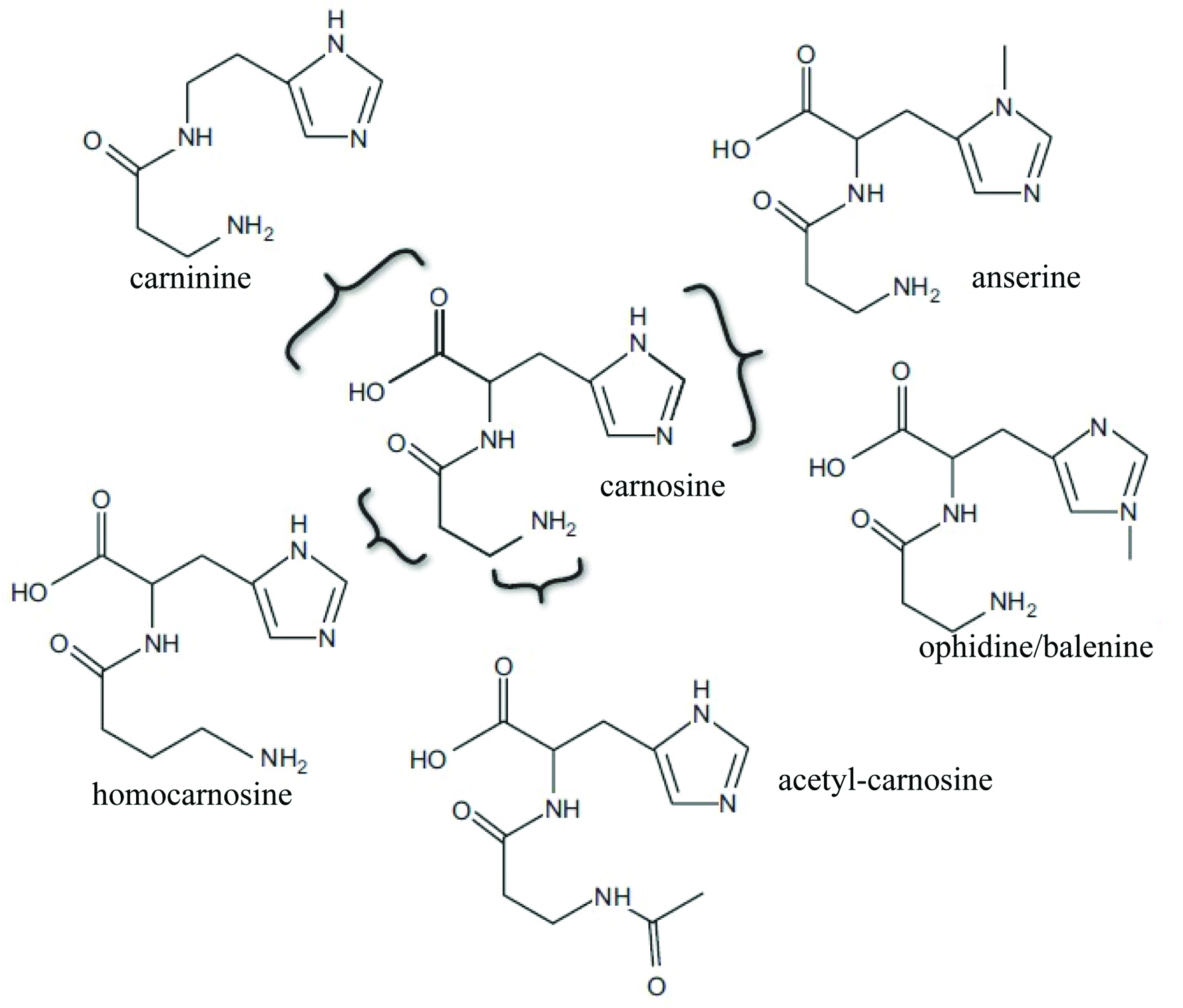
Figure 1. Chemical structure of carnosine (β-alanine-L-histidine) and its naturally occurring derivatives (adapted from Boldyrev et al., 2013).
| Journal of Food Bioactives, ISSN 2637-8752 print, 2637-8779 online |
| Journal website www.isnff-jfb.com |
Review
Volume 5, March 2019, pages 8-17
Carnosine—a natural bioactive dipeptide: bioaccessibility, bioavailability and health benefits
Figures
Tables
| Type of extract | Carnosine concentration (mg/g) | Reference |
|---|---|---|
| Beef | 5.8∼7.12 | Marcolini et al., 2015 |
| Pork | 0.13∼4.19 | Mora et al., 2008 |
| Horse | 1.22∼4.07 | Abe, 2000 |
| Lamb | 7.06 | Jones et al., 2011 |
| Rabbit | 3.6∼4.6 | Peiretti and Meineri, 2015 |
| Chicken | 0.66∼1.83 | Jung et al., 2013 |
| Turkey | 0.86∼7.9 | Gil-Agustí et al., 2008, Peiretti et al., 2012 |
| Duck | 0.14∼0.16 | Lee et al., 2015 |
| Lobster | 0.1 | Kantha et al., 2000 |
| Prawn | 9.25∼11.6 | Preedy, 2015 |
| Tuna | 5.29 | Jones et al., 2011 |
| Mackerel | 7.78 | Jones et al., 2011 |
| Rainbow Trout | 0.11 | Jones et al., 2011 |
| Bonito (katsuo) | 5.0∼7.9 | Kantha et al.,2000 |
| Conger eel (hamo) | 0.2 | Kantha et al., 2000 |
| Bioactive activity | Experimental Model | Observed effects | Reference |
|---|---|---|---|
| Anti-oxidant | Chemical detection | Inactivate free radicals;Metal-chelating, such as copper;Conjugating with potentially toxic aldehydic lipid oxidation products | Decker et al., 2000 |
| Cerebellum granule cells | ↓ROS signal;↓Excitotoxic effect of N-methyl-d-aspartic acid (NMDA) | Boldyrev et al., 2004 | |
| Rat | ↓Lactate dehydrogenase | Park et al., 2005 | |
| Wistar rats and Mongolian gerbils | ↓Lipid peroxidation of brain membrane; ↑Resistance of neuronal membranes under oxidation; ↓malonyl dialdehyde (MDA) | Dobrota et al., 2005 | |
| Chemical detection | Metal-chelating;↓Lipid peroxidation | Derave et al., 2010 | |
| Colon cancer cells | ↓ROS; ↓ERK1/2 phosphorylation; ↓Proliferation | Iovine et al., 2012 | |
| Sprague-Dawley male rats | ↓Oxidative stress; Restores the histopathological and biochemical signs; ↓Apoptotic condition | Aydin et al., 2015 | |
| Mice | ↑Glutathione peroxidase (GPX), Superoxide dismutase (SOD), and Catalase;↓ MDA ; ↓Reactive oxygen species | Tsai et al., 2010 | |
| Recreationally-active men volunteer | ↑GSH ↑CAT, PRX2, SOD1, and TRX1 | Varanoske et al., 2017 | |
| Broiler chickens | ↑Antioxidant enzymes; ↓MDA and carbonyl compounds | Cong et al., 2017 | |
| Wistar rats | ↑CAT,SOD and total antioxidant capacity (TAC) | Hasanein and Felegari, 2017 | |
| Cirrhotic rats | ↑Locomotor activity; ↓ROS formation; ↓Liver TBARS; ↑GSH in liver; ↓ Protein carbonylation; ↓Liver fibrogenesis; ↓ Liverfailure | Jamshidzadeh et al., 2017 | |
| Human erythrocytes and lymphocytes | ↓Oxidative damage; Restores enzyme activities and antioxidant power | Husain and Mahmood, 2017 | |
| Anti-glycation | Chemical detection | ↑Glyc3P-induced loss of enzyme activity; ↓Protein modification | Seidler, 2000 |
| Human Peritoneal Mesothelial Cells (HPMC) | ↑HPMC viability; ↓Advanced glycation end products (AGE) complications; Protection cellular protein from modification; | Alhamdani et al., 2007 | |
| E. coli K-12 Strains | ↑Cell viability;↓Glycation; ↓Methylglyoxal toxicity | Pepper et al., 2010 | |
| Gray chinchilla rabbits | ↓Gycation-induced diabetic cataract | Babizhayev et al., 2012 | |
| Human lens epithelial cell models | ↓Glycosylated damage; ↑Lens cell viability | Abdelkader et al., 2016 | |
| Aged rats | ↓AGE and MDA; ↓Protein carbonyl and advanced oxidized protein products | Bingül et al., 2017 | |
| Anti-inflammatory | Caco-2 cells | ↓IL-8 secretion | Son et al., 2008 |
| Male Balb/cA mice | ↓c-reactive protein (CRP), IL-6 tumor and TNF-α | Liu et al., 2008 | |
| Balb/cA mice | ↓IL-6, TNF-α, and MCP-1 | Yan et al., 2009 | |
| Mice | ↓TNF-α and IL-6 levels | Tsai et al., 2010 | |
| Elderly people | ↓MCP-1 and IL-8 | Hisatsune et al., 2016 | |
| Sepsis-induced male albino rats. | ↓IL-β, TNF-α, and IL-8;↑IκBα; ↓p65 and p-IKKα/β (Ser 180/Ser 181); ↓Lung Injury | Sun et al., 2017 | |
| Human cancer cell line HepG2 | ↓PI3K and Akt ; ↑Caspase-8 ; ↓Proliferation; ↑apoptosis | Liu et al., 2017 | |
| Wistar rats | ↓TNF-α and IL-6 levels | Hasanein and Felegari, 2017 |